The Promise of Deep Learning in Medical Imaging
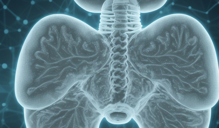
In the high-stakes world of medical diagnostics, clarity is paramount. Magnetic Resonance Imaging (MRI) stands as a cornerstone for visualizing the intricate structures of the human brain, aiding in the detection of tumors, aneurysms, and a host of neurological disorders. However, inherent limitations such as noise and resolution constraints often obscure critical details, hindering accurate diagnoses. Now, a new frontier is emerging: deep learning models, particularly Convolutional Neural Networks (CNNs), are being engineered to overcome these challenges, promising to sharpen MRI images and unlock unprecedented diagnostic utility.
This article delves into the architecture, implementation, and ethical considerations of building such a model, offering a comprehensive guide for researchers and practitioners alike. The potential impact is transformative, poised to enhance diagnostic accuracy, improve patient outcomes, and redefine the landscape of medical imaging. The application of AI, particularly deep learning, in medical imaging shares conceptual similarities with its use in other complex domains, such as weather prediction and environmental modeling, where extracting meaningful insights from noisy and high-dimensional data is crucial.
For example, just as CNNs can denoise MRI images, similar architectures are used to refine weather models by filtering out atmospheric noise and improving the resolution of forecasts. The core principles of feature extraction and pattern recognition remain consistent, highlighting the versatility of deep learning across diverse scientific fields. Furthermore, the evolution of AI language models, moving beyond simple text generation to sophisticated reasoning and contextual understanding, mirrors the advancements in medical image analysis. The ability of models like U-Net and ResNet to not only enhance image quality but also to identify subtle anomalies reflects a broader trend in AI towards more nuanced and context-aware applications.
This convergence underscores the potential for cross-pollination of ideas and techniques, driving innovation across various disciplines. The development of AI-driven MRI enhancement tools also raises important ethical considerations, mirroring debates surrounding the use of AI in other sensitive areas. Ensuring fairness, transparency, and accountability is paramount to prevent biases in the training data from perpetuating health disparities. As deep learning models become more integrated into medical workflows, it is crucial to establish clear guidelines and regulatory frameworks to govern their use and protect patient privacy.
This necessitates a collaborative effort involving researchers, clinicians, policymakers, and ethicists to navigate the complex ethical landscape and ensure that AI is used responsibly and equitably in medical imaging and beyond. The availability of powerful deep learning frameworks like TensorFlow and PyTorch has significantly accelerated the development and deployment of MRI enhancement models. These platforms provide researchers with the necessary tools to design, train, and evaluate complex neural networks, fostering innovation and collaboration. Moreover, the increasing availability of large, annotated medical image datasets is fueling the progress of AI in radiology. As the field continues to evolve, we can expect to see even more sophisticated AI-powered tools that not only enhance image quality but also assist radiologists in making more accurate and timely diagnoses, ultimately improving patient care.
Data Acquisition and Preprocessing: Laying the Foundation
The journey to enhanced MRI clarity begins with meticulous data acquisition and preprocessing. MRI data, unlike standard images, often suffers from unique artifacts such as bias fields – subtle, spatially varying intensity distortions. Addressing these requires specialized techniques like N4 bias field correction, an algorithm designed to estimate and remove these unwanted variations. Normalization is equally crucial, ensuring that image intensities fall within a consistent range, typically between 0 and 1. This step mitigates the impact of varying scanner settings and patient-specific factors.
Data augmentation techniques, such as rotations, translations, and slight intensity variations, are also employed to artificially expand the training dataset, improving the model’s robustness and generalization capabilities. According to a recent study published in *Radiology*, proper preprocessing can significantly improve the performance of deep learning models in medical image analysis. The National Institutes of Health (NIH) emphasizes the importance of standardized data acquisition protocols to ensure the reliability and reproducibility of AI-driven medical imaging solutions.
Extending beyond basic image adjustments, preprocessing in the context of deep learning for medical image enhancement, particularly with convolutional neural networks (CNNs), mirrors techniques used in other domains like weather prediction. For instance, just as atmospheric data undergoes rigorous cleaning and scaling before being fed into forecasting models, MRI data requires similar treatment to optimize the performance of CNN architectures like U-Net or ResNet. The presence of noise in MRI scans can be likened to atmospheric turbulence, hindering accurate analysis.
Denoising algorithms, therefore, play a crucial role, analogous to how signal processing techniques filter out noise in weather radar data. Data imputation, used to handle missing values in environmental modeling, also finds its parallel in MRI processing when dealing with incomplete scans. The selection of preprocessing techniques also impacts the effectiveness of AI language models used in radiology report generation. If the input MRI data is poorly preprocessed, the resulting radiology reports generated by AI may contain inaccuracies or misinterpretations.
This highlights the interconnectedness of different AI modalities within the medical field. Furthermore, the success of super-resolution techniques, which aim to enhance the resolution of MRI images, heavily relies on high-quality preprocessing. A blurry or distorted input will inevitably lead to a suboptimal super-resolved output, regardless of the sophistication of the deep learning model. Frameworks like TensorFlow and PyTorch provide extensive tools for implementing these preprocessing steps, but the expertise lies in understanding the nuances of MRI data and tailoring the pipeline accordingly.
Considering ethical implications, standardized data acquisition and preprocessing are essential for mitigating biases in AI-driven medical diagnostics. If the training data disproportionately represents a specific demographic or scanner type, the resulting deep learning model may exhibit skewed performance across different patient populations. This concern echoes similar challenges in AI-powered environmental monitoring, where biases in sensor data can lead to inaccurate predictions and unfair resource allocation. Therefore, adhering to FAIR (Findable, Accessible, Interoperable, Reusable) data principles and employing robust validation techniques are crucial for ensuring the equitable and reliable deployment of AI in medical imaging, promoting trust and widespread adoption of these technologies in radiology and beyond.
CNN Architecture and Implementation: Building the Engine
The architecture of the CNN is the engine driving image enhancement. U-Net, with its distinctive encoder-decoder structure and skip connections, has emerged as a favorite for medical image segmentation and enhancement. The encoder progressively downsamples the input image, capturing high-level features, while the decoder reconstructs the image at a higher resolution. Skip connections bridge corresponding layers in the encoder and decoder, allowing the model to retain fine-grained details often lost during downsampling. ResNet, another popular choice, employs residual connections to mitigate the vanishing gradient problem, enabling the training of deeper and more complex networks.
For MRI enhancement, a hybrid approach combining elements of U-Net and ResNet may offer optimal performance. Consider a U-Net architecture with ResNet blocks in the encoder path. This allows for capturing both global context and fine-grained details, crucial for denoising and super-resolution in magnetic resonance imaging (MRI). Implementation in TensorFlow or PyTorch is straightforward. Here’s a simplified PyTorch snippet for a convolutional block: python import torch.nn as nn class ConvBlock(nn.Module): def __init__(self, in_channels, out_channels): super(ConvBlock, self).__init__() self.conv = nn.Conv2d(in_channels, out_channels, kernel_size=3, padding=1) self.relu = nn.ReLU(inplace=True) def forward(self, x): return self.relu(self.conv(x))
Custom loss functions are essential for guiding the model’s learning process. Perceptual loss, which measures the difference between high-level image features extracted by a pre-trained CNN, encourages the model to generate visually appealing results. Structural Similarity Index (SSIM) loss penalizes distortions in image structure. A combination of L1 loss, SSIM loss, and perceptual loss often yields the best results. The code for SSIM loss can be implemented using libraries such as `pytorch_msssim`. According to Dr.
Fei-Fei Li, a leading AI researcher at Stanford University, ‘The choice of architecture and loss function is critical for achieving state-of-the-art performance in medical image enhancement.’ The evolution of CNN architectures for medical image enhancement mirrors advancements seen in AI language models. Just as transformer networks superseded recurrent neural networks (RNNs) in natural language processing, novel CNN designs are constantly emerging to address the specific challenges of MRI data. For instance, attention mechanisms, inspired by their success in language models like ChatGPT, are now being integrated into CNNs for medical imaging.
These mechanisms allow the network to focus on the most relevant features in the MRI scan, improving denoising and super-resolution capabilities. This adaptation highlights the cross-pollination of ideas between different domains of artificial intelligence (AI). The selection of an appropriate loss function is paramount for training effective deep learning models for MRI enhancement. Beyond perceptual loss and SSIM, researchers are exploring adversarial loss functions, commonly used in generative adversarial networks (GANs). In this context, the CNN acts as a generator, attempting to produce enhanced MRI images that are indistinguishable from real, high-quality images.
A discriminator network then tries to differentiate between the generated and real images. This adversarial training process can lead to more realistic and visually appealing results. Furthermore, the principles of transfer learning, prevalent in both AI language models and machine learning for weather prediction, can be applied by fine-tuning pre-trained CNNs on large datasets of natural images for the specific task of MRI enhancement, significantly reducing training time and improving performance. Considering the computational demands of training deep learning models, especially for 3D MRI data, efficient implementation is crucial.
Frameworks like TensorFlow and PyTorch offer tools for distributed training, allowing researchers to leverage multiple GPUs or even entire clusters of machines. Techniques like mixed-precision training, which uses a combination of 16-bit and 32-bit floating-point numbers, can significantly reduce memory consumption and accelerate training without sacrificing accuracy. Furthermore, the deployment of these models in clinical settings requires careful consideration of computational resources. Model compression techniques, such as pruning and quantization, can reduce the size and complexity of the CNN, making it feasible to run on resource-constrained devices commonly found in radiology departments and improving the speed and efficiency of medical diagnostics.
Training Strategies: Optimizing Performance
Training deep learning models for MRI enhancement demands a multifaceted approach, extending beyond basic hyperparameter tuning to encompass advanced strategies that leverage insights from diverse AI applications. Data augmentation remains a cornerstone, but its implementation should be tailored to the specific characteristics of MRI data. For example, simulating variations in patient positioning or scanner calibration can generate more robust training sets. Furthermore, techniques borrowed from AI language models, such as adversarial training, can be adapted to enhance the model’s ability to distinguish between genuine anatomical features and noise artifacts.
Consider a generative adversarial network (GAN) where one network generates realistic MRI images and another tries to distinguish them from real images, leading to improved denoising capabilities. This approach mirrors how language models refine their understanding of context through adversarial interactions. The FDA emphasizes the need for rigorous validation and testing of AI-based medical devices to ensure safety and efficacy. According to a report by the World Health Organization (WHO), AI has the potential to improve access to healthcare in underserved communities, but it is essential to address ethical considerations and potential biases.
The complexities of training algorithms for medical image enhancement are considerable, but the potential benefits for improved diagnostics are immense. Hyperparameter optimization for convolutional neural networks (CNNs) used in medical image enhancement requires a strategic approach, often guided by insights from machine learning in weather prediction and predictive environmental modeling. Just as weather models benefit from ensemble methods and adaptive learning rates, MRI enhancement models can gain from similar techniques. Bayesian optimization, which intelligently explores the hyperparameter space based on past performance, is particularly effective.
For instance, the optimal learning rate for a U-Net architecture used for MRI super-resolution might be influenced by the specific characteristics of the input data, such as signal-to-noise ratio. Techniques like transfer learning, where models pre-trained on large datasets of natural images or even synthetic MRI data, can accelerate training and improve performance, especially when the amount of real MRI data is limited. A ResNet architecture, known for its ability to handle vanishing gradients in very deep networks, may benefit from pre-training on a related medical imaging task, such as CT scan segmentation, before fine-tuning on MRI data.
This approach reduces the demand for large, labeled datasets, a common bottleneck in medical image analysis. Beyond conventional metrics like PSNR and SSIM, the evaluation of medical image enhancement models should incorporate perceptual loss functions that better reflect human visual perception. Drawing inspiration from AI language models that strive for coherent and contextually relevant outputs, perceptual loss functions aim to minimize the difference between the enhanced MRI image and the ground truth image in terms of high-level features extracted by a pre-trained CNN.
This approach can lead to more visually appealing and clinically relevant enhancements, even if the PSNR values are not significantly improved. Furthermore, explainable AI (XAI) techniques are becoming increasingly important in radiology, allowing clinicians to understand why a particular enhancement was generated and to assess its reliability. Techniques like Grad-CAM can highlight the regions of the input image that most strongly influenced the model’s output, providing valuable insights into the model’s decision-making process. The application of AI in medical image enhancement holds immense promise for improving diagnostic accuracy and patient outcomes, but it requires careful consideration of both technical and ethical challenges.
The choice of deep learning framework, such as TensorFlow or PyTorch, also plays a crucial role in the development and deployment of MRI enhancement models. PyTorch, with its dynamic computational graph and ease of debugging, is often favored for research and experimentation, while TensorFlow, with its robust production capabilities and support for various hardware platforms, is well-suited for deployment in clinical settings. The integration of these models into existing radiology workflows requires seamless compatibility with picture archiving and communication systems (PACS) and adherence to established DICOM standards.
Furthermore, the development of AI-powered tools for medical diagnostics raises important ethical considerations, including data privacy, algorithmic bias, and the potential for job displacement. Addressing these challenges requires a collaborative effort involving researchers, clinicians, regulators, and patients to ensure that AI is used responsibly and ethically to improve healthcare for all. The future of medical image enhancement lies in the development of more sophisticated and robust AI algorithms that can seamlessly integrate into clinical practice and improve the accuracy and efficiency of medical diagnostics.
Evaluation, Ethical Considerations, and Future Directions
Evaluating the performance of an MRI enhancement model demands a multifaceted approach, blending quantitative metrics with the nuanced qualitative assessments of experienced radiologists. While Peak Signal-to-Noise Ratio (PSNR) and Structural Similarity Index (SSIM) offer valuable quantitative insights—PSNR gauging the ratio of signal strength to noise and SSIM assessing perceived image quality changes—they often fall short of capturing subtle, yet clinically significant, improvements crucial for accurate medical diagnostics. These metrics, while useful, are analogous to evaluating a weather forecasting model solely on temperature accuracy, neglecting critical factors like precipitation intensity or wind patterns.
Therefore, the subjective expertise of radiologists remains indispensable. Their ability to discern improved clarity, reduced noise artifacts, and the preservation of fine anatomical details in enhanced images is paramount for validating the clinical utility of deep learning models. Real-world case studies serve as compelling evidence of a model’s effectiveness in enhancing diagnostic accuracy and fostering trust in AI-driven solutions. For instance, research published in *Nature Medicine* showcased a deep learning model’s ability to enhance the detection of small brain lesions in MRI scans, leading to earlier diagnoses and more effective treatment interventions.
This is akin to AI language models moving beyond simple text generation to performing complex reasoning and problem-solving tasks, showcasing capabilities beyond initial expectations. The success hinges not only on the architecture of the convolutional neural networks (CNNs) employed, such as U-Net or ResNet implemented using TensorFlow or PyTorch, but also on the sophisticated training strategies, including the use of perceptual loss functions designed to mimic human perception of image quality, thereby optimizing for clinically relevant features rather than just numerical scores.
Future research should explore innovative deep learning architectures, such as transformers, which have demonstrated remarkable capabilities in natural language processing and are now making inroads into medical imaging, potentially revolutionizing medical image enhancement and denoising. Furthermore, integrating multi-modal imaging data, like PET and CT scans, could provide a more comprehensive view of the patient’s condition, leading to more accurate diagnoses and personalized treatment plans. This mirrors the trend in AI language models towards multi-modal understanding, where models can process and integrate information from various sources, such as text, images, and audio.
However, addressing ethical considerations and potential biases is paramount. AI models can inadvertently perpetuate or amplify existing biases present in the training data, leading to disparities in healthcare outcomes. Careful attention must be paid to data diversity and fairness to ensure equitable access to the benefits of AI-driven medical image enhancement. As artificial intelligence (AI) continues to transform magnetic resonance imaging (MRI) and medical image enhancement, a collaborative ecosystem involving researchers, clinicians, policymakers, and even experts in fields like machine learning in weather prediction (given the shared challenges of dealing with noisy, high-dimensional data) is essential.
This collaboration will ensure that these technologies are developed and deployed responsibly and ethically, ultimately benefiting patients worldwide. The ongoing evolution of neural network architectures, pushing beyond large language models and towards more specialized and efficient designs, will be crucial for realizing the full potential of deep learning in medical diagnostics. Only through such concerted efforts can we harness the power of AI to improve the clarity and accuracy of MRI, leading to better patient care and outcomes.